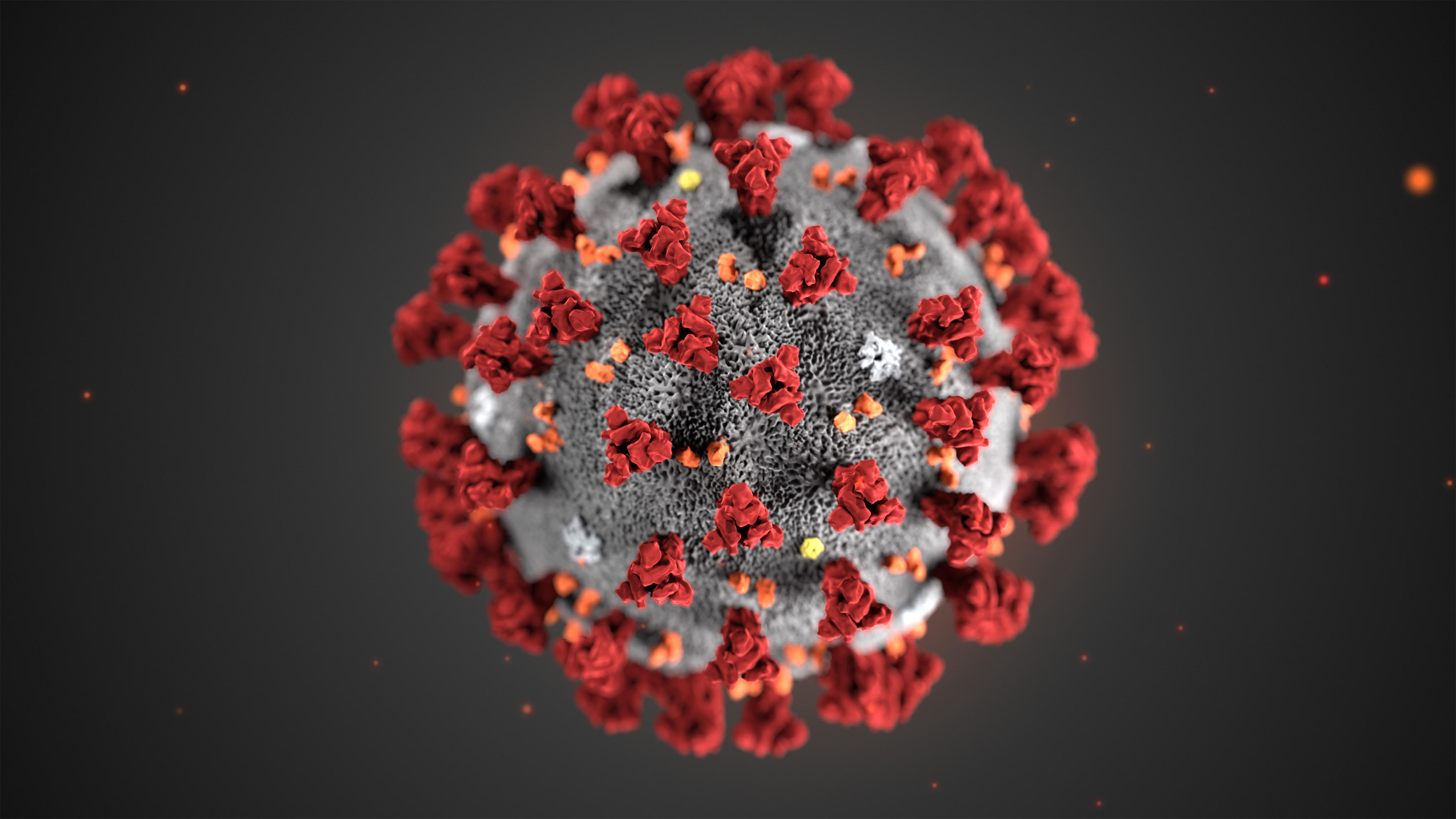
Novel Coronavirus (COVID-19)
Learn about novel coronavirus (COVID-19), our preparedness and updated policies to protect our community, patients, and staff. Learn more here.
Emergency
570-253-8141
Patient Room:
570-253-8609
When prompted enter 3-digit room number
Human Resources
570-253-8995
Wayne Memorial Hospital
601 Park Street
Honesdale, PA 18431
Click the map below for turn by turn directions.
At Wayne Memorial we have one of the best-equipped radiology departments in Northeastern Pennsylvania with ultrasound, nuclear medicine, digital and traditional mammography, bone density testing, CT Scan, PET/CT Scan, X-ray, MRI. 3D Mammography and, as of 2024, interventional radiology.
Wayne Memorial Hospital – (570) 253-8124
601 Park Street, Honesdale, PA 18431
24 hour service. Services offered: X-rays, MRI, CT, Pet Scans, Nuclear Medicine, Digital Mammograms, Ultrasound, Bone Density, Vascular Studies.
Wayne Memorial Outpatient Services – Carbondale – (570) 282 – 1637
Carbondale Family Health Center, 150 Brooklyn St., Carbondale, 18407
Services offered: X-ray and EKG, Monday – Friday, 7:00 am – 3:30 pm
Wayne Memorial Outpatient Services – Hamlin – (570) 689-4670
Hamlin Family Health Center, 543 Easton Turnpike – Route 191, Hamlin, PA 18427
Services offered: X-ray Monday through Friday 7:30am – 6:30pm (possibly later some days, call ahead), Saturday and Sunday 9 am – 3 pm; ultrasounds Monday, Wednesday and Thursday by appointment only from 8:30 am to 5 p.m.
Wayne Memorial Outpatient Services – Pike County – (570) 775-4278
Pike Family Health Center, 750 Route 739, Lords Valley, PA 18428 | Monday – Friday 7:30am- 4 pm
Services offered: X-rays, 3D Digital Mammograms, Bone Density Testing
Rayus Radiology, one of the top ranked radiology groups in the United States, provides professional interpretations and consultations with sub-specialization experts available 24 hours a day, 7 days a week, 365 days a year.
Mammography – Our digital mammography unit in the Women’s Imaging Center at Wayne Memorial Hospital and our highly trained staff are fully accredited by the American College of Radiology. We offer 3D mammography via the GE Senographe Pristina system, at the hospital, in our Pike County Medical Center and in our mobile unit.
The 3D Pristina mammography equipment from GE is designed for safety, comfort and clarity. This digital breast tomosynthesis system (DBT) reconstructs a 3-dimensional image from a single scan and delivers “superior diagnostic accuracy at the same dose as 2D mammography, the lowest patient dose of all government approved DBT systems,” according to GE. Moreover, with rounded edges, a thinner receptor plate and armrests instead of hand grips, the Pristina reduces much of the discomfort of earlier mammography systems.
Mobile Mammography – The 3D system is also in our mobile unit, which travels to Carbondale, Hamlin, Forest City and Lake Como.
To make an appointment for mammography, with a prescription from your provider, call 570-251-6689.
Ultrasound – Our three new Siemens Acuson Antares Ultrasound Systems deliver superb image quality and allow for a full range of fast, non-invasive diagnoses. Some clinical applications for this imaging technology include:
Diagnostic Section (X-RAY) – General Electric XRD Revolution equipment allows for direct digital capture of x-ray images. Each image is available for review within 3-5 seconds, allowing for maximum efficiency in diagnosis and treatment. Digital radiographic/fluoroscopic equipment and computed radiography units linked to our picture archiving and communications system, allow for immediate access to x-ray studies anywhere within the healthcare system.
Bone Density Testing Section – Our new Hologic QDR 4500 DEXA-Scan unit offers the most accurate diagnosis and screening of osteoporosis and other bone disorders.
Nuclear Medicine – Two gamma cameras, an ADAC Vertex Plus Dual-Head SPECT Gamma Camera and an ADAC ACR 3000 with Pegasus 20 Technology use minimal radiation to provide the most accurate diagnoses of conditions involving the heart, lungs, biliary system, gall bladder, kidneys, thyroid and bones.
Computerized Axial Tomography (CAT SCAN) – Our newly acquired state-of-the-art Aquilion PRIME from Toshiba is capable of producing 160 images or “slices” per rotation. It provides faster exams at the lowest doses of radiation that are reasonably achievable while producing high-quality images for precise diagnoses.
The speed of the Aquilion PRIME allows clinicians to obtain critical patient information faster than before. Shorter exams mean less contrast and radiation are required, making the procedure much safer. The equipment also offers more comfort for patients and expands the range of patients who can be imaged—from pediatric to bariatric– partly because of the Aquilion PRIME’s large bore opening and the industry’s widest couch.
Some typical procedures performed include:
Magnetic Resonance Imaging (MRI) – A GE 1.5 Tesla, with Excite High Definition Technology provides a quantum improvement in the speed and resolution of MRI studies. These improved studies allow doctors to diagnose a wide range of conditions including:
This technology also allows radiologists to perform breast biopsies with MRI guidance.
PET/CT Scans—positron emission tomography with computer-assisted tomography. This imaging procedure, used to determine the location and stage of cancerous tissue, can help prevent unnecessary surgery, biopsies and inappropriate treatments. PET/CT has a major impact on the clinical evaluations of cancer patients, and in many cases will enable physicians to begin treatment earlier and increase the odds for successful patient outcomes.
The precise images obtained with PET/CT are not available with other technologies, such as X-ray, MRI, or CT alone. The PET/CT equipment combines or “fuses” the images of a PET scanner (metabolic function of cells) with the CT scanner (anatomic location of body structures) into one extremely detailed image.
Interventional Radiology — specialized physician service. Imaging procedures such as fluoroscopy and X-rays are used to help guide procedures, including biopsies and angiograms, to treat various blood vessel and lymphatic conditions.